Automatic 3D Cardiovascular MR Segmentation with
Densely-Connected Volumetric ConvNets
Lequan Yu1 Jie-Zhi Cheng2 Qi Dou1 Xin Yang1 Hao Chen1 Jing Qin3 Pheng Ann Heng1
1The Chinese Univeristy of Hong Kong 2Chang Gung University 3The Hong Kong Polytechnic University
Abstract
Automatic and accurate whole-heart and great vessel segmentation from 3D cardiac magnetic resonance (MR) images plays an important role in the computer-assisted diagnosis and treatment of cardiovascular disease. However, this task is very challenging due to ambiguous cardiac borders and large anatomical variations among different subjects. In this paper, we propose a novel densely-connected volumetric convolutional neural network, referred as DenseVoxNet, to automatically segment the cardiac and vascular structures from 3D cardiac MR images. The DenseVoxNet adopts the 3D fully convolutional architecture for effective volume-to-volume prediction. From the learning perspective, our DenseVoxNet has three compelling advantages. First, it preserves the maximum information flow between layers by a densely-connected mechanism and hence eases the network training. Second, it avoids learning redundant feature maps by encouraging feature reuse and hence requires fewer parameters to achieve high performance, which is essential for medical applications with limited training data. Third, we add auxiliary side paths to strengthen the gradient propagation and stabilize the learning process. We demonstrate the effectiveness of DenseVoxNet by comparing it with the state-of-the-art approaches from HVSMR 2016 challenge in conjunction with MICCAI, and our network achieves the best dice coefficient. We also show that our network can achieve better performance than other 3D ConvNets but with fewer parameters.
Method
 |
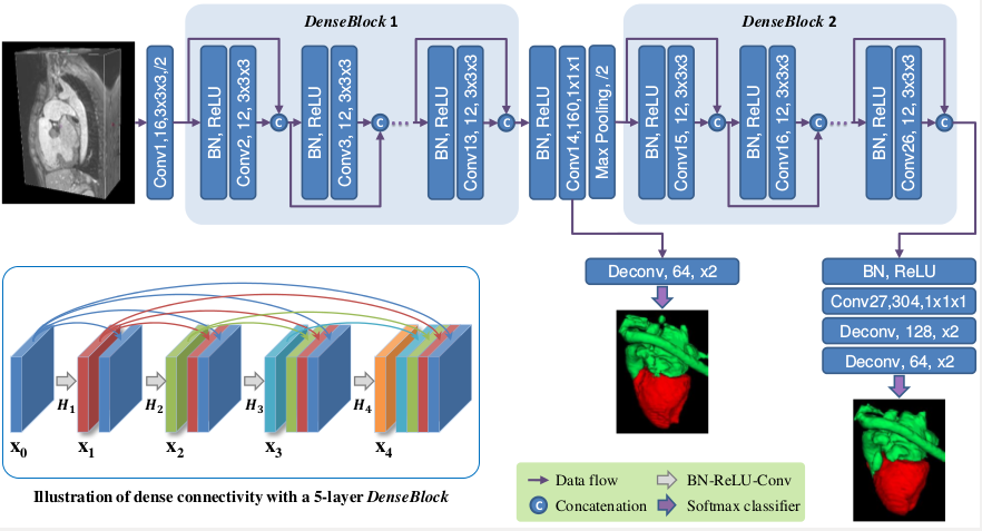
The architecture of our DenseVoxNet. It consists of two DenseBlocks and all operations are implemented in a 3D manner. The green and red color denotes the output of blood pool and myocardium. The graph in left bottom illustrates the dense connectivity scheme taking a 5-layer DenseBlock as an example. |
Results

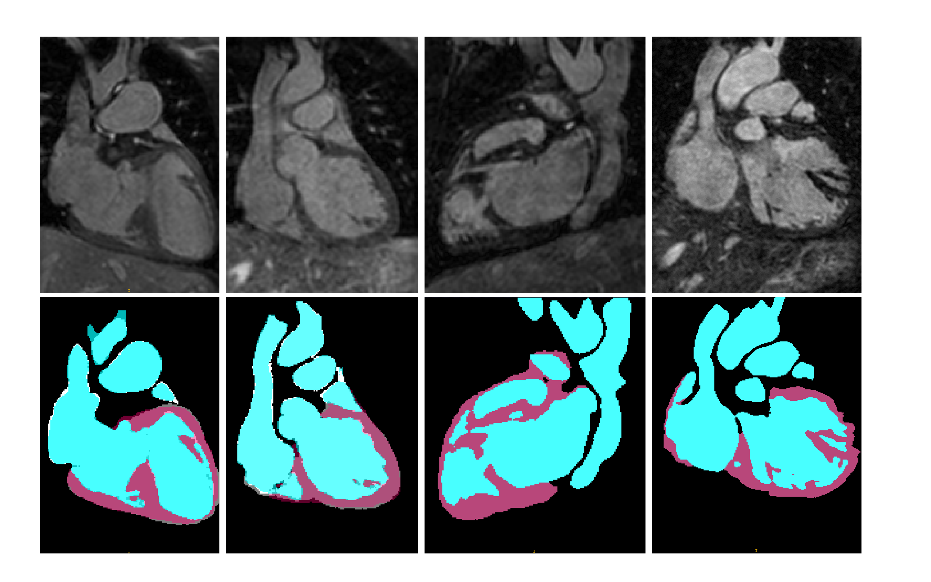
Segmentation results on training images (the first two) and testing images (the last two). The blue and purple color denotes our segmentation results for blood pool and myocardium, respectively, and segmentation ground truth is also presented in white and gray regions in the first two samples. |

|
||

|
Downloads

|
"Automatic 3D Cardiovascular MR Segmentation with Densely-Connected Volumetric ConvNets", MICCAI 2017 Lequan Yu, Jie-Zhi Cheng, Qi Dou, Xin Yang, Hao Chen, Jing Qin, Pheng Ann Heng |
Last update: Dec., 2016